Science to watch in 2023 according to Nature
 Credit: Adobe Stock
The journal Nature presented seven breakthrough technologies that can accelerate the development of science in 2023. In addition to the Webb Telescope or the CRISPR system, the list also includes embryo cultivation from stem cells, a project by Professor Magdalena Żernicka-Goetz.
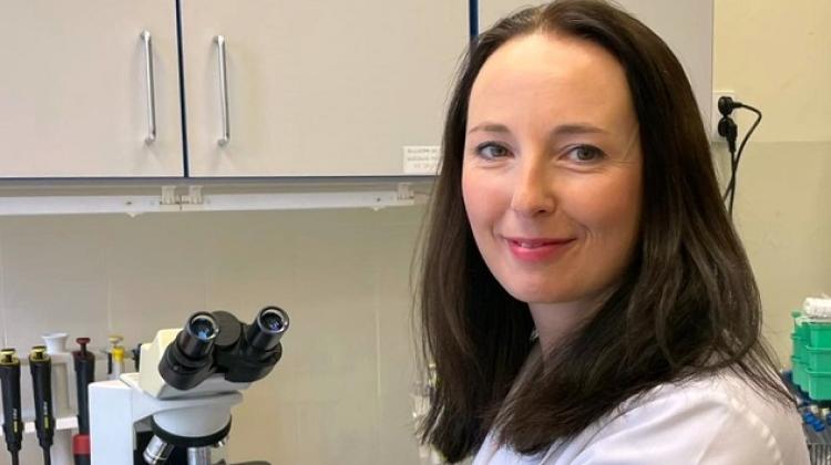
Last year, Professor Magdalena Żernicka-Goetz's team from the University of Cambridge grew an artificial mouse embryo that contained an active, beating heart and a brain bud. Instead of reproductive cells, the researchers used three types of stem cells. The embryo survived seven days. After that, it stopped developing due to defects, but the achievement may be the key to understanding exactly how individual organs form, and to predicting the possible errors that may occur.
Professor Żernicka-Goetz told the BBC: “This period of human life is so mysterious, so to be able to see how it happens in a dish - to have access to these individual stem cells, to understand why so many pregnancies fail and how we might be able to prevent that from happening - is quite special.”
She and her colleagues are working to extend the life of the embryo and are even considering working on human embryos. Although this is associated with strong controversies, there are indications that the gate to learning how a human being is created may soon open.
The work of the team with the participation of the Polish researcher is one of the seven achievements presented by 'Nature' as technologies to watch in 2023.
Among the potential breakthroughs in the field of biology, Nature also lists single-cell metabolomics. The metabolome is a set of all substances that participate in the body's metabolism - fats, sugars, proteins and many molecules of other types. So far, it has been possible to analyse this system in relation to tissues or organs, but it may soon become possible for single cells. Scientists believe that many cells may look identical, but differ precisely in terms of metabolism. The metabolome is the active component of the cell. Understanding the metabolism of single cells can help to understand numerous diseases. Such studies are already being carried out, but they are extremely expensive. Meanwhile, a team from the European Molecular Biology Laboratory in Heidelberg, Germany has developed a much cheaper method, based on the use of light microscopy, standard analytical methods and special computer software. Scientists predict the creation of a metabolic atlas that will show what is happening in various cells.
Another aspect of cells is the proteome, which represents proteins produced by the body, tissue, or cell. They also carry invaluable information about a possible disease or correct biochemical processes. Just like in the case of DNA researchers analyse the sequence of the four basic 'letters' of the genetic code, in the case of proteins, they analyse the amino acid sequence. DNA can be copied many times, which significantly facilitates research. This is not possible with proteins.
According to Nature's expert Michael Eisenstein, this means that the molecules detected cannot always be identified unambiguously, and low-abundance proteins in a mixture are often overlooked. Now, however, new horizons are opening up thanks to single-molecule protein sequencing, in which, as the name suggests, even a single protein molecule is sufficient for analysis.
One such method is being developed at the University of Texas at Austin. In this process, individual amino acids are fluorescently labelled and then sheared off one by one from the end of a surface-coupled protein as a camera captures the resulting fluorescent signal. A somewhat similar method was developed at Quantum-Si, a biotech company that wants to start selling its devices this year. So the inside of cells should soon begin to reveal all its secrets to biologists.
The same may be true of tissues thanks to volume electron microscopy. In a typical approach, it can take 200 sections to cover the volume of just a single cell with an electron microscope. The new technique may herald a revolution - thanks to it, it is possible to obtain images of tissue samples encompassing many cubic millimetres. This makes it easier to observe the inside of cells, as reported by scientists in pioneering papers. This method may change extremely expensive and painstaking brain research, conducted on the scale of individual cells and neuronal connections.
At the same time, scientists are getting better and better at manipulating cells, for example with the now famous CRISPR editing, which opened the way to precise genetic engineering, including the development of gene therapies. However, as the author of the summary in Nature explains, conventional CRISPR enzymes read anywhere between 1percent and 10 percent of the genome. This is a major limitation that may soon be overcome. A team from Massachusetts General Hospital in Boston has created new enzyme variants that can read practically the entire genome. Their downside is that they are less accurate, but researchers hope that they will be able to solve this problem. Moreover, because the CRISPR system is derived from bacteria, there are also many different variants in nature. Biologists analyse them in search of the best versions. A group from the University of Trento has already found a set of Cas9 variants, which they estimate could collectively target more than 98 percent of known disease-causing mutations in humans.
While some look into the micro world, others turn their attention to space. Nature highlights the importance of the long-awaited James Webb Telescope (JWST). Last year, the instrument showed its unique capabilities, for example by capturing signatures from a galaxy formed 200 million years after the Big Bang, observing young stars, interstellar dust, distant planets and the Solar System - and even weather phenomena on Titan. Thanks to infrared detectors, JWST can see what the Hubble Telescope could not, and generate images with much higher resolution. In the coming months, scientists expect more cosmic revelations.
Others look to the past. Last year, specialists from the University of Groningen in the Netherlands used radiocarbon dating to obtain a date accuracy of at least one year. During the study of Viking travels in Newfoundland (Canada), they were able to determine that one of the trees was cut down in 1021, probably in the spring. The typical method of measuring traces of the radioactive isotope carbon-14 is usually precise only to within a couple of decades.
The new dating method is based on the 2012 discovery by a Japanese team that dated a distinctive spike in carbon-14 levels in the rings of a Japanese cedar tree to 774-5 CE. A similar spike was later confirmed in trees elsewhere in the world. Later, researchers identified other such spikes dating as far back as 7176 BCE. They are probably related to solar storms. It is enough to count the number of rings - starting from the ring with this type of spike - to determine the year of felling of a given tree. Based on the thickness of the last ring, it is possible to deduce the time of year when a tree was felled. Numerous other events can be deduced from this information. The team from Groningen hopes to use this method to the Mayan empire. The scientists believe that they will have absolute records for ancient civilizations to the exact year. (PAP)
mat/ zan/ kap/
tr. RL
Przed dodaniem komentarza prosimy o zapoznanie z Regulaminem forum serwisu Nauka w Polsce.


